41 the human heart and its labels
Heart Labeling Quiz: How Much You Know About Heart Labeling? Here is a Heart labeling quiz for you. The human heart is a vital organ for every human. The more healthy your heart is, the longer the chances you have of surviving, so you better take care of it. Take the following quiz to know how much you know about your heart. Questions and Answers 1. What is #1? 2. What is #2? 3. What is #3? 4. What is #4? Heart Anatomy: Labeled Diagram, Structures, & Function 24 Feb 2022 — Anatomy of the heart study guide PDF. Includes labeled diagrams, along with the location and function of structures including the atria, ...
The Anatomy of the Heart, Its Structures, and Functions The heart is the organ that helps supply blood and oxygen to all parts of the body. It is divided by a partition (or septum) into two halves. The halves are, in turn, divided into four chambers. The heart is situated within the chest cavity and surrounded by a fluid-filled sac called the pericardium. This amazing muscle produces electrical ...
The human heart and its labels
Parts Of The Human Heart | Science Trends The parts of the human heart can be broken down into four chambers, muscular walls, vessels, and a conductive system. The two upper chambers are called the atria, with lower parts called ventricles. These all work together to make up the vital function of your heart. Everybody knows that the human heart is the essential organ in our bodies. Human Heart - Diagram and Anatomy of the Heart - Innerbody The heart is a muscular organ about the size of a closed fist that functions as the body's circulatory pump. It takes in deoxygenated blood through the veins and delivers it to the lungs for oxygenation before pumping it into the various arteries (which provide oxygen and nutrients to body tissues by transporting the blood throughout the body). Human Heart - Anatomy and Functions | Location and Chambers Anatomy and Functions of the Human Heart. The human heart is the organ that pumps blood throughout the body via the vessels of the circulatory system, supplying oxygen and nutrients to the tissues and removing carbon dioxide and other wastes. Pumping the blood through the arteries, capillaries, and veins is the major function of the heart.
The human heart and its labels. Label the Heart Diagram | Quizlet Start studying Label the Heart. Learn vocabulary, terms, and more with flashcards, games, and other study tools. Heart: Anatomy and Function - Cleveland Clinic Your heart is the main organ of your cardiovascular system, a network of blood vessels that pumps blood throughout your body. It also works with other body systems to control your heart rate and blood pressure. Your family history, personal health history and lifestyle all affect how well your heart works. Appointments 800.659.7822 File:Diagram of the human heart (no labels).svg - Wikimedia Commons This file is licensed under the Creative Commons Attribution-Share Alike 4.0 International license.: You are free: to share - to copy, distribute and transmit the work; to remix - to adapt the work; Under the following conditions: attribution - You must give appropriate credit, provide a link to the license, and indicate if changes were made. You may do so in any reasonable manner, but ... Human Heart Labeling Teaching Resources | Teachers Pay Teachers Human Heart Parts and Blood Flow Labeling Worksheets - Diagram/Graphic Organizer by TechCheck Lessons 22 $2.25 Zip This resource contains 2 worksheets for students to (1) label the parts of the human heart and (2) Fill in a flowchart tracing the path of blood flowing though the circulatory system. Answer keys included.
Labelling the heart — Science Learning Hub Labelling the heart — Science Learning Hub Labelling the heart Add to collection The heart is a muscular organ that pumps blood through the blood vessels of the circulatory system. Blood transports oxygen and nutrients to the body. It is also involved in the removal of metabolic wastes. Topics Concepts Citizen science Teacher PLD Glossary Sign in File:Diagram of the human heart (cropped).svg - Wikipedia File:Diagram of the human heart (cropped).svg. Size of this PNG preview of this SVG file: 611 × 600 pixels. Other resolutions: 244 × 240 pixels | 489 × 480 pixels | 782 × 768 pixels | 1,043 × 1,024 pixels | 2,086 × 2,048 pixels | 663 × 651 pixels. This is a file from the Wikimedia Commons. Information from its description page there is ... Heart - Wikipedia The human heart is situated in the mediastinum, at the level of thoracic vertebrae T5-T8.A double-membraned sac called the pericardium surrounds the heart and attaches to the mediastinum. The back surface of the heart lies near the vertebral column, and the front surface sits behind the sternum and rib cartilages. The upper part of the heart is the attachment point for several large blood ... Diagram of Human Heart and Blood Circulation in It Exterior of the Human Heart A heart diagram labeled will provide plenty of information about the structure of your heart, including the wall of your heart. The wall of the heart has three different layers, such as the Myocardium, the Epicardium, and the Endocardium. Here's more about these three layers. Epicardium
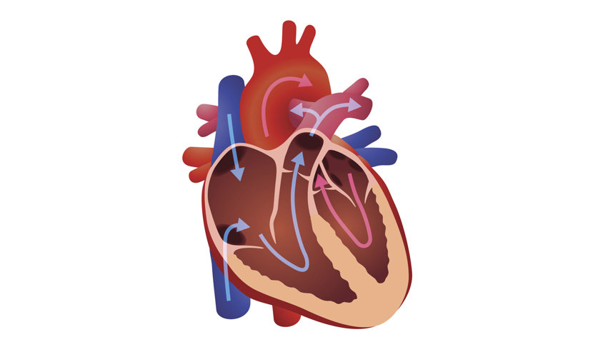
File:Diagram of the human heart (cropped).svg - Wikimedia ... Diagram of the human heart, created by Wapcaplet in Sodipodi. ... No labels version ... Added shadows. Left main pulmonary artery with its first division. How to draw internal structure of Human heart - Easy version Internal structure of human heart shows four chambers viz. two atria and two ventricles and couple of blood vessels opening into them. The wall of two ... A Diagram of the Heart and Its Functioning Explained in Detail The heart blood flow diagram (flowchart) given below will help you to understand the pathway of blood through the heart.Initial five points denotes impure or deoxygenated blood and the last five points denotes pure or oxygenated blood. 1.Different Parts of the Body ↓ 2.Major Veins ↓ 3.Right Atrium ↓ 4.Right Ventricle ↓ 5.Pulmonary Artery ↓ 6.Lungs Human Heart - Anatomy, Functions and Facts about Heart The human heart is divided into four chambers, namely two ventricles and two atria. The ventricles are the chambers that pump blood and atrium are the chambers that receive the blood. Among which, the right atrium and ventricle make up the "right portion of the heart", and the left atrium and ventricle make up the "left portion of the heart." 5.
Our World | What is the heart?
Heart Diagram with Labels and Detailed Explanation - BYJUS Diagram of Heart. The human heart is the most crucial organ of the human body. It pumps blood from the heart to different parts of the body and back to the heart. The most common heart attack symptoms or warning signs are chest pain, breathlessness, nausea, sweating etc. The diagram of heart is beneficial for Class 10 and 12 and is frequently ...
Sampoerna Wallpaper: The Heart Diagram Labeled
Human heart: Anatomy, function & facts | Live Science The human heart has four chambers: two upper chambers (the atria) and two lower ones (the ventricles), according to the National Institutes of Health. The right atrium and right ventricle together...

Parts Of Heart Diagram Stock Illustration - Download Image Now - iStock
Human Heart for Kids: 2 Fun Heart Models plus Worksheets We also make a playdough heart model and completed the free printable heart worksheets. This is just one of our human body activities as part of our human body for kids unit studying anatomy for grade 1, grade 2, grade 33, grade 4, grade 5, grade 6, and garde 7 students. Whether you are a parent, teacher, or homeschooler you will love these ...

Human Nervous System Structure and Functions Explained With Diagrams - Bodytomy
Human Heart Diagram Labeled | Science Trends Human Heart Diagram Labeled Daniel Nelson 1, January 2019 | Last Updated: 3, March 2020 The human heart is an organ responsible for pumping blood through the body, moving the blood (which carries valuable oxygen) to all the tissues in the body. Without the heart, the tissues couldn't get the oxygen they need and would die.

Congestive Heart Failure: The Essence of Heart Failure Course | CEUfast Nursing Continuing Education
13+ Heart Diagram Templates - Sample, Example, Format Download Human heart is a complicated figure and for students from science, they will often need the images of the heart for its illustration. The above collection of heart samples will make it easier for students to download, print and use it in their projects. The images with labels and detailed explanations can also be used in text books.

Simplified Heart Labeled Decal | Shop Fathead Anatomical Images Graphics
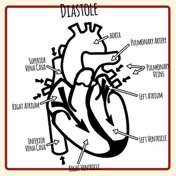
Label the heart — Science Learning Hub Label the heart Interactive Add to collection In this interactive, you can label parts of the human heart. Drag and drop the text labels onto the boxes next to the diagram. Selecting or hovering over a box will highlight each area in the diagram. Right ventricle Right atrium Left atrium Pulmonary artery Left ventricle Pulmonary vein Semilunar valve
32 Label This Anterior View Of The Human Heart - Labels Design Ideas 2020
Human Heart Diagram - Human Body Pictures - Science for Kids Find free pictures, photos, diagrams, images and information related to the human body right here at Science Kids. Photo name: Human Heart Diagram Picture category: Human Body Image size: 70 KB Dimensions: 600 x 600 Photo description: This is an excellent human heart diagram which uses different colors to show different parts and also labels a number of important heart component such as the ...

New Photos in Anatomy of human body organs
Human heart with labelling Images, Stock Photos & Vectors Find Human heart with labelling stock images in HD and millions of other royalty-free stock photos, illustrations and vectors in the Shutterstock collection ...

30 Human Heart With Label - Labels Design Ideas 2020
147 Heart Anatomy With Labels Premium High Res Photos - Getty Images Browse 147 heart anatomy with labels stock photos and images available, or start a new search to explore more stock photos and images. of 3. NEXT.
How the human heart did become associated with love? | 3D ORGANON
Human Heart (Anatomy): Diagram, Function, Chambers, Location in Body The heart is a muscular organ about the size of a fist, located just behind and slightly left of the breastbone. The heart pumps blood through the network of arteries and veins called the...

Business Diary: October 2011
PDF Analyzing the Human Heart - Beyond the Classroom working. Its job is to pump blood to the lungs and to all of the body tissues. In this activity you will use a diagram of the heart to analyze the way in which the heart works. l. Using the following word list, label the various parts of the heart on the diagram. Right ventricle Left venfficle Upper vena cava Lower vena cava Aorta
:max_bytes(150000):strip_icc()/human-heart-circulatory-system-598167278-5c48d4d2c9e77c0001a577d4.jpg)
The Anatomy of the Heart, Its Structures, and Functions
How to Draw a Human Heart: 11 Steps (with Pictures) - wikiHow If you're trying to identify parts of the heart for a class you're taking, it's good practice to draw the heart yourself and label each segment. You can refer to your textbook in order to label the: Aorta Superior vena cava Inferior vena cava Right and left atria Right and left ventricles Pulmonary veins and arteries 5

Pokemon Cosplay: Kawaii Pokemon Pikachu Cosplay Couple
Simple heart diagram labeled - Anatomy - Pinterest We provide you a simple heart diagram to draw and learn. Simple heart diagram labeled with accurate labels. Most frequent question in exam to draw human heart ...
Human Heart - Label the Diagram Anatomy Clip Art Set Commercial Use
Anatomy of a Human Heart - uofmhealth Located between the lungs in the middle of the chest, the heart pumps blood through the network of arteries and veins known as the cardiovascular system. It pushes blood to the body's organs, tissues and cells. Blood delivers oxygen and nutrients to every cell and removes the carbon dioxide and other waste products made by those cells.

Something That Won't Stop Until You Die
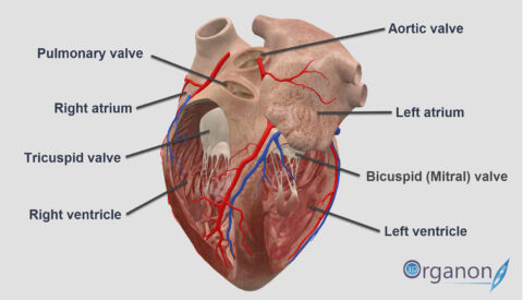
A Labeled Diagram of the Human Heart You Really Need to See The human heart, comprises four chambers: right atrium, left atrium, right ventricle and left ventricle. The two upper chambers are called the left and the right atria, and the two lower chambers are known as the left and the right ventricles. The two atria and ventricles are separated from each other by a muscle wall called 'septum'.

Human Heart Pictures with Labels Best Of File Diagram Of the Human Heart Hug Wikimedia Mons ...
The 18 parts of the human heart, and their functions 9. Left ventricle. The left ventricle contains the strongest muscles in the whole heart. From this ventricle, blood is pumped into the aortic artery, which divides to water the rest of the body's blood. The blood pressure generated by this ventricle must be much higher than that generated by the right ventricle. 10.
Post a Comment for "41 the human heart and its labels"